เทคโนโลยีการรักษา
MRI Brain Scan
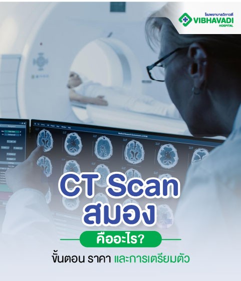
Advanced Diagnostic Imaging for Brain and Neurological Health What Is an MRI Brain Scan? An MRI brain scan (Magnetic Resonance Imaging of the brain) is a non-invasive imaging test that uses powerful magnetic fields and radio waves to create detailed images of the brain and surrounding nervous system structures. It is widely used for diagnosing brain tumors, strokes, aneurysms, multiple sclerosis (MS), brain injuries, and other neurological disorders. Unlike CT scans or X-rays, an MRI does not use ionizing radiation, making it a safer option for many patients, especially for repeated imaging and for younger individuals. Why Get a Brain MRI? A brain MRI provides in-depth information that helps neurologists and radiologists: Detect brain tumors or cysts Identify stroke-related damage Diagnose multiple sclerosis or epilepsy Evaluate causes of chronic headaches or migraines Investigate memory loss, confusion, or seizures Monitor brain inflammation or infections Examine brain structure after trauma Who Should Consider an MRI of the Brain? Recommended for People Who Have: Persistent or severe headaches Dizziness or vision problems Seizures or unexplained neurological symptoms History of brain injury or stroke Unexplained behavioral or memory changes Suspected brain tumor, aneurysm, or MS Family history of neurological diseases Brain MRI Procedure: Step-by-Step Before the Scan Medical Screening Inform the medical team if you have metal implants, a pacemaker, or are pregnant. Fill out a safety questionnaire. Change into a Gown Metal-free clothing is required. Jewelry, watches, hairpins, or anything metallic must be removed. During the Scan Positioning You will lie flat on a cushioned table that slides into the MRI scanner (a tunnel-shaped device). Noise & Communication The machine makes loud tapping or thumping sounds during scanning. You will wear earplugs or headphones and can communicate with the technician at any time. Stillness Required Staying still is important to ensure high-quality images. Depending on the study, a contrast agent may be injected into a vein to enhance visibility of specific brain areas. After the Scan You can resume normal activities immediately. If contrast was used, drink water to help flush it from your system. Results will typically be reviewed by a radiologist and sent to your referring doctor. Duration of Brain MRI Without contrast: Approx. 30–45 minutes With contrast: Approx. 45–60 minutes Total time including preparation: ~1 hour Preparation for a Brain MRI Things to Do Before the Exam: Inform your doctor about metal implants, kidney issues (if contrast is needed), and pregnancy Fasting: May be required if contrast will be used No caffeine: Avoid caffeine 2–3 hours before the exam if you’re prone to anxiety or claustrophobia Take prescribed medication: As directed by your physician Tips for Claustrophobic Patients: Vibhavadi offers comfort-focused MRI suites Sedation or mild anti-anxiety medication may be available with physician approval MRI Brain Price and Packages at Vibhavadi Hospital We offer transparent, competitive pricing with no hidden costs. MRI Brain without contrast: Starting from 9,000 THB MRI Brain with contrast: Starting from 13,000 THB Package includes radiologist interpretation and medical report Pricing may vary based on your clinical condition and physician recommendation Safety of MRI Brain Scans MRI is a safe and well-established procedure No radiation is involved Contrast agents are generally safe, with rare allergic reactions Not suitable for patients with certain metal implants or pacemakers Related Services at Vibhavadi Hospital Neurology Clinic and Consultations CT Scan (Brain, Spine, and other regions) EEG (Electroencephalogram) Stroke Assessment and Management Dementia and Alzheimer’s Screening Emergency Brain Injury Evaluation Annual Check-up Packages with Imaging Add-ons Insurance and Health Benefits Direct billing available with partnered insurance providers Accepted under most health insurance plans (inpatient & outpatient) Packages eligible for use with annual check-up benefits or corporate health programs Ask our staff for a pre-authorization letter if needed Book Your Appointment Scheduling your MRI brain scan is easy: Website: https://www.vibhavadi.com/th Call Center: Contact Radiology Department or International Service Desk Walk-In: Available but subject to slot availability Our MRI Technology Vibhavadi Hospital uses high-field MRI scanners (1.5T and 3.0T), providing: High-resolution imaging Shorter scan times Enhanced comfort for patients Dedicated software for brain analysis, tumor detection, and stroke evaluation Our Medical Team Our radiology department is staffed by: Board-certified radiologists Neurologists with expertise in neuroimaging MRI technicians trained in patient safety and scan optimization Multilingual staff for international patients Frequently Asked Questions (FAQ) Is an MRI brain scan painful? No. It is completely non-invasive and painless. Can I eat before my MRI scan? Yes, unless you are having contrast — in that case, you may need to fast for 4–6 hours. How long before I get the results? Typically, the radiologist's report is available within 24–48 hours. Urgent cases are prioritized. Is MRI safe for children? Yes, MRI is safe for children. Pediatric cases may require sedation depending on age and cooperation. Will I need contrast? That depends on what your doctor is investigating. For tumor evaluation, infection, or vascular issues, contrast is often recommended.
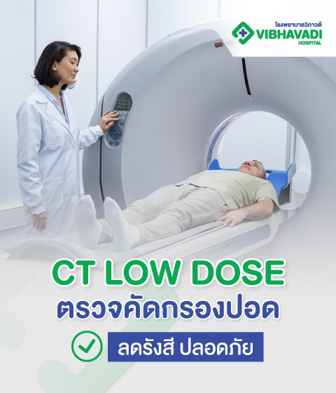
CT Low Dose
CT Low Dose Lung Screening at Vibhavadi Hospital Early Detection. Low Radiation. High Precision. What Is a CT Low Dose? A CT Low Dose (Low-Dose Computed Tomography, or LDCT) is a medical imaging technique that uses significantly less radiation than a conventional CT scan. It is specifically designed for lung cancer screening and early detection of other pulmonary diseases. Unlike chest X-rays, CT low dose provides detailed cross-sectional images of the lungs, allowing doctors to detect abnormalities at an early stage — often before symptoms appear. Why Choose Low-Dose CT? Reduced Radiation Exposure One of the key benefits of CT low dose is that it uses up to 90% less radiation than standard CT scans while still delivering high-quality images. This is especially important for: Routine screening Long-term monitoring High-risk individuals (e.g., smokers or those with occupational exposure) Effective for Early Lung Cancer Detection Research shows that LDCT can reduce lung cancer mortality by 20% in high-risk populations. The scan can detect: Early-stage tumors Lung nodules Chronic lung diseases (e.g., emphysema, fibrosis) Infection-related changes Who Should Get a CT Low Dose Lung Scan? High-Risk Individuals LDCT lung screening is recommended for people who are: Aged 50–80 Current or former smokers (20 pack-years or more) Have chronic respiratory symptoms (e.g., persistent cough, shortness of breath) Family history of lung cancer Previous exposure to asbestos, radon, or industrial pollutants People With These Symptoms Should Consider Screening: Chronic cough lasting more than 3 weeks Unexplained weight loss Fatigue or wheezing Coughing up blood Repeated respiratory infections How the CT Low Dose Scan Works Overview of the Procedure Low-dose CT uses X-ray technology to create detailed 3D images of your lungs. The scan takes only a few seconds, and the entire process is quick, painless, and non-invasive. Step-by-Step CT Scan Process Check-In and Briefing You will be greeted by the radiology staff and given instructions. No contrast dye is needed for standard LDCT lung screening. Preparation and Positioning You will lie down on the scanning table. The radiology technician will ensure you are in the correct position — usually lying on your back with arms raised above your head. Scanning The table will move through the CT scanner, capturing multiple images in just a few seconds while you hold your breath briefly. Completion and Review The scan is reviewed by a radiologist, and a report is sent to your physician or discussed during your follow-up. How Long Does It Take? Total time: 10–15 minutes Actual scan: less than 30 seconds No recovery time needed Preparation for CT Low Dose Before the Scan Wear comfortable, metal-free clothing Remove jewelry or objects containing metal No fasting or medication changes required (unless advised) After the Scan Resume daily activities immediately No side effects or recovery period Your doctor will contact you with results or schedule a follow-up Advantages of CT Low Dose Scanning Safe and low radiation exposure Highly effective in detecting early-stage lung cancer Quick and painless Non-invasive No use of contrast (unless specified) Detects other conditions such as COPD or pulmonary fibrosis Why Choose Vibhavadi Hospital for CT Low Dose? Advanced Imaging Technology At Vibhavadi Hospital, we use state-of-the-art low-dose CT scanners that ensure the best balance of image clarity and minimal radiation. Our equipment is regularly calibrated to meet international safety standards. Expert Radiologists Our radiology team includes board-certified radiologists with experience in thoracic imaging and lung cancer screening. They provide accurate interpretation and detailed reports. Comprehensive Care Pathway If an abnormality is detected, we offer: Pulmonary specialist referral Lung function testing Bronchoscopy or biopsy services Oncology consultation Related Services at Vibhavadi Hospital Chest X-ray and MRI Pulmonary function test (PFT) Lung cancer screening packages Smoking cessation clinic Health check-up programs Internal medicine and respiratory care Frequently Asked Questions (FAQ) Is CT Low Dose safe? Yes. It uses significantly less radiation than standard CT scans and is considered safe for routine lung screening. Is contrast dye needed? Not for standard lung cancer screening. Contrast may be used for other types of CT scans. How often should I get a CT Low Dose? If you're in a high-risk group (e.g., smoker or ex-smoker), annual screening is recommended — especially for ages 50–80. Will it detect other lung issues? Yes. It can detect emphysema, fibrosis, infections, and even signs of tuberculosis or COVID-19-related changes. Can I drive after the scan? Yes. There are no sedatives or recovery periods involved.
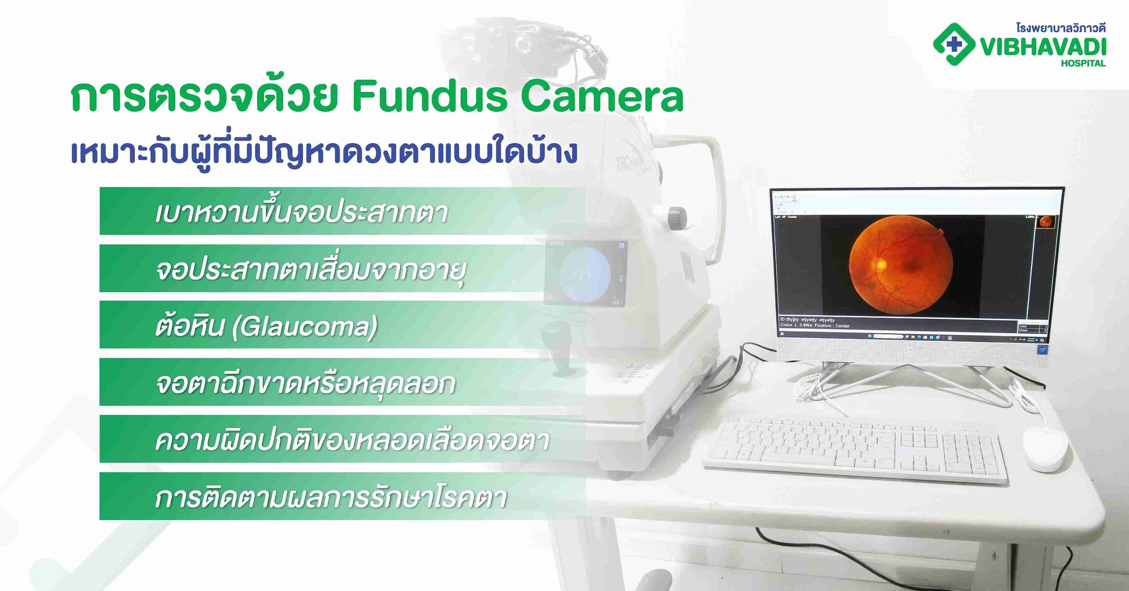
Fundus Camera
Fundus Camera Eye Checkup at Vibhavadi Hospital Early Detection. Clear Vision. Peace of Mind. A fundus camera eye checkup is a non-invasive diagnostic procedure that captures high-resolution images of the interior surface of the eye, including the retina, optic disc, macula, and posterior pole. At Vibhavadi Hospital, we provide advanced retinal imaging services using state-of-the-art fundus camera technology. This allows for early detection of vision-threatening diseases such as diabetic retinopathy, glaucoma, macular degeneration, and hypertensive retinopathy. What Is a Fundus Camera? A fundus camera is a specialized low-power microscope with an attached camera designed to photograph the interior surface of the eye (the fundus). The images it captures are used by ophthalmologists to detect and monitor a wide range of eye conditions and systemic diseases that affect the retina and optic nerve. Why Retinal Imaging Matters The Importance of the Retina The retina is a light-sensitive tissue lining the back of the eye. It plays a critical role in vision by converting light into neural signals sent to the brain. Retinal damage is often irreversible and can lead to partial or complete vision loss if not detected early. Silent Progression of Eye Diseases Many serious eye diseases develop without noticeable symptoms in the early stages. Regular retinal imaging allows for: Early diagnosis Timely intervention Monitoring disease progression Preventing vision loss Who Should Get a Fundus Camera Eye Checkup? High-Risk Groups The following individuals are strongly recommended to undergo a fundus camera eye checkup: Diabetics (Type 1 and Type 2) Hypertensive patients Individuals over 40 years old Smokers and heavy alcohol consumers People with a family history of glaucoma, AMD, or retinal disorders Patients with frequent migraines or visual aura Individuals with high cholesterol or cardiovascular risk People With These Symptoms Should Also Consider Screening: Blurred or distorted vision Difficulty seeing at night Flashes of light or floaters Sudden vision loss Eye pain or pressure Common Conditions Detected by Fundus Photography Diabetic Retinopathy A common complication of diabetes, this condition damages the blood vessels in the retina and can lead to blindness if untreated. Glaucoma Increased intraocular pressure can damage the optic nerve, causing gradual vision loss. Age-related Macular Degeneration (AMD) This affects central vision, often in adults over 60, and is a leading cause of blindness. Hypertensive Retinopathy High blood pressure can cause structural damage to the retina, leading to blurred vision or even bleeding within the eye. Retinal Detachment or Tears These require urgent care and can be visually assessed with fundus imaging. How the Fundus Camera Procedure Works Step-by-Step Process Pupil Dilation You may be given eye drops to dilate your pupils for better visibility of the retina. Image Capture You will rest your chin on a support while the camera takes images of the back of your eye. Non-Invasive and Quick The procedure is painless, takes only 5–10 minutes, and requires no downtime. No Preparation Required There is no special preparation needed. However, if dilation drops are used, you should avoid driving for a few hours post-exam. Benefits of Fundus Camera Eye Checkup Non-invasive and painless Quick and efficient Highly detailed imaging for accurate diagnosis Can be stored and used for future comparison Supports early intervention in eye and systemic diseases Vibhavadi Hospital's Retinal Imaging Services Our Advanced Fundus Camera Technology We use high-resolution digital fundus cameras capable of wide-angle and detailed imaging. Our equipment enables: Fluorescein angiography (optional) Optical coherence tomography (OCT) integration Automated and manual capture modes Secure image storage for tracking patient history Professional Interpretation by Expert Ophthalmologists Every scan is reviewed by our board-certified ophthalmologists, trained in both retinal diseases and systemic conditions visible through fundus imaging. Detailed reports and recommendations are provided promptly. Seamless Referral for Further Care If the scan reveals abnormalities, patients are referred to specialists such as: Retina specialists Glaucoma specialists Internal medicine or endocrinology (for diabetes or hypertension) Related Services at Vibhavadi Hospital Comprehensive eye exams Diabetic eye screening Glaucoma screening and management Macular degeneration treatment OCT (Optical Coherence Tomography) IOP (Intraocular Pressure) testing LASIK and ICL surgery consultation Booking a Fundus Camera Checkup How to Make an Appointment You can schedule your checkup via: Website: https://www.vibhavadi.com/th/package/fundus-camera-eye-checkup Call Center: Contact the Eye Center for available slots Walk-in: Visit our Ophthalmology Department on-site Clinic Hours Monday–Friday: 8:00 AM – 5:00 PM Saturday: 9:00 AM – 12:00 PM Closed on Sundays and public holidays Cost and Coverage Checkup Package Price The Fundus Camera Eye Checkup at Vibhavadi Hospital is currently offered at: Only 600 THB (subject to hospital promotions) This includes: Fundus image capture Expert interpretation Preliminary consultation Insurance and Health Benefits Covered under some corporate health screening programs Tax-deductible as health checkup expense in Thailand May be claimable through private insurance (check with provider) Meet Our Medical Team Our Ophthalmology Team includes: Retinal and macular disease specialists Diabetic eye care experts Glaucoma management professionals Experienced technicians trained in advanced imaging systems Each member is committed to high-standard, patient-centered care. Frequently Asked Questions (FAQ) Is the fundus camera procedure safe? Yes. It is a non-invasive, completely safe procedure that uses light and digital imaging—not X-rays or radiation. Will it hurt? Not at all. The procedure is quick and painless, although dilation drops might cause temporary light sensitivity. Do I need this checkup even if I have no vision problems? Yes. Many retinal conditions, especially diabetic and hypertensive retinopathy, do not show symptoms until advanced stages. How often should I get this checkup? Diabetics: Annually or as advised Individuals over 40: Every 1–2 years Anyone with vision symptoms: Immediately Can children have a fundus camera exam? Yes, particularly if they have systemic diseases or symptoms. Pediatric retinal screening is available upon request.
