Fundus Camera Eye Examination | Vibhavadi
Fundus Camera Eye Checkup at Vibhavadi Hospital
Early Detection. Clear Vision. Peace of Mind.
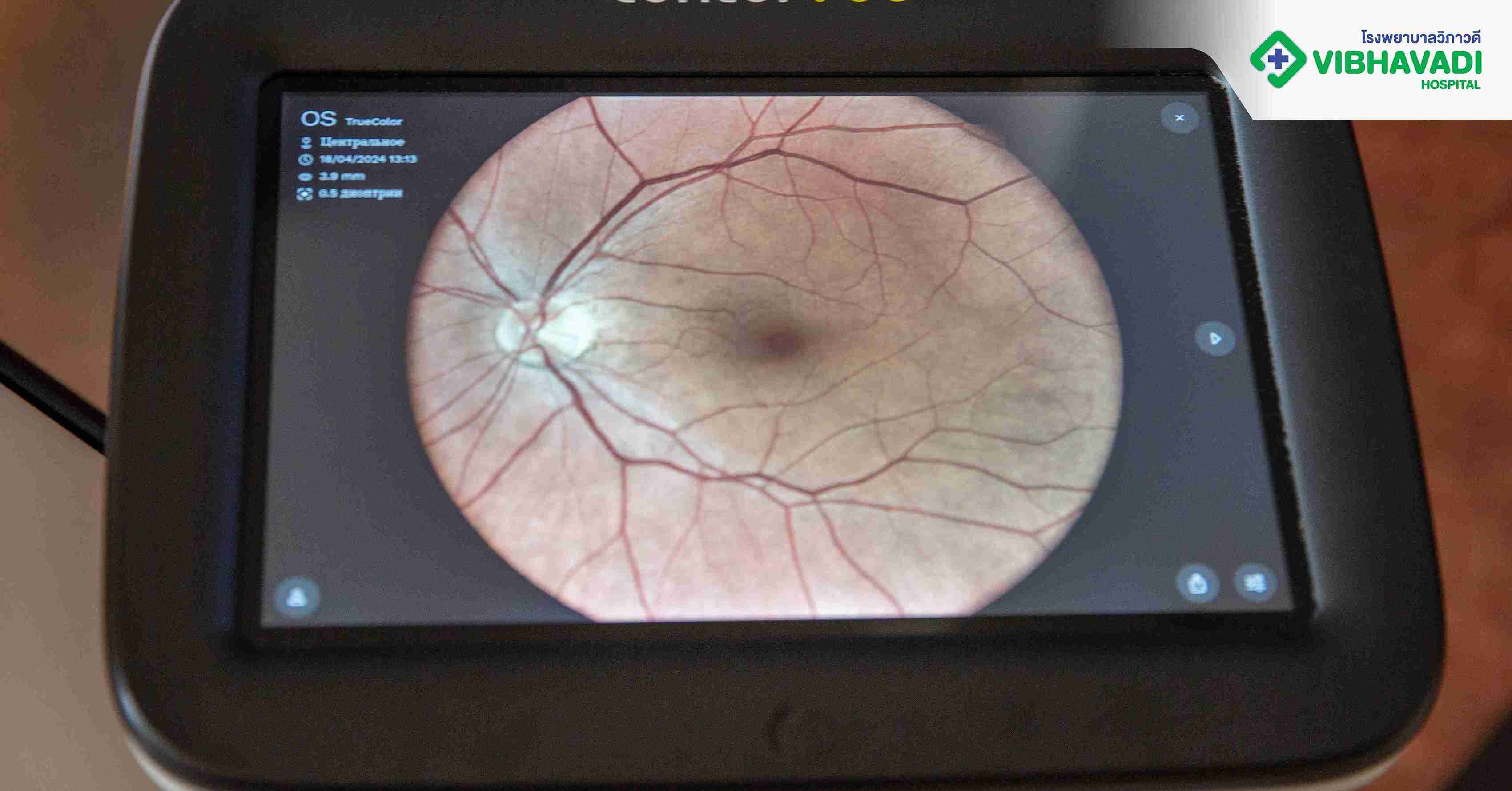
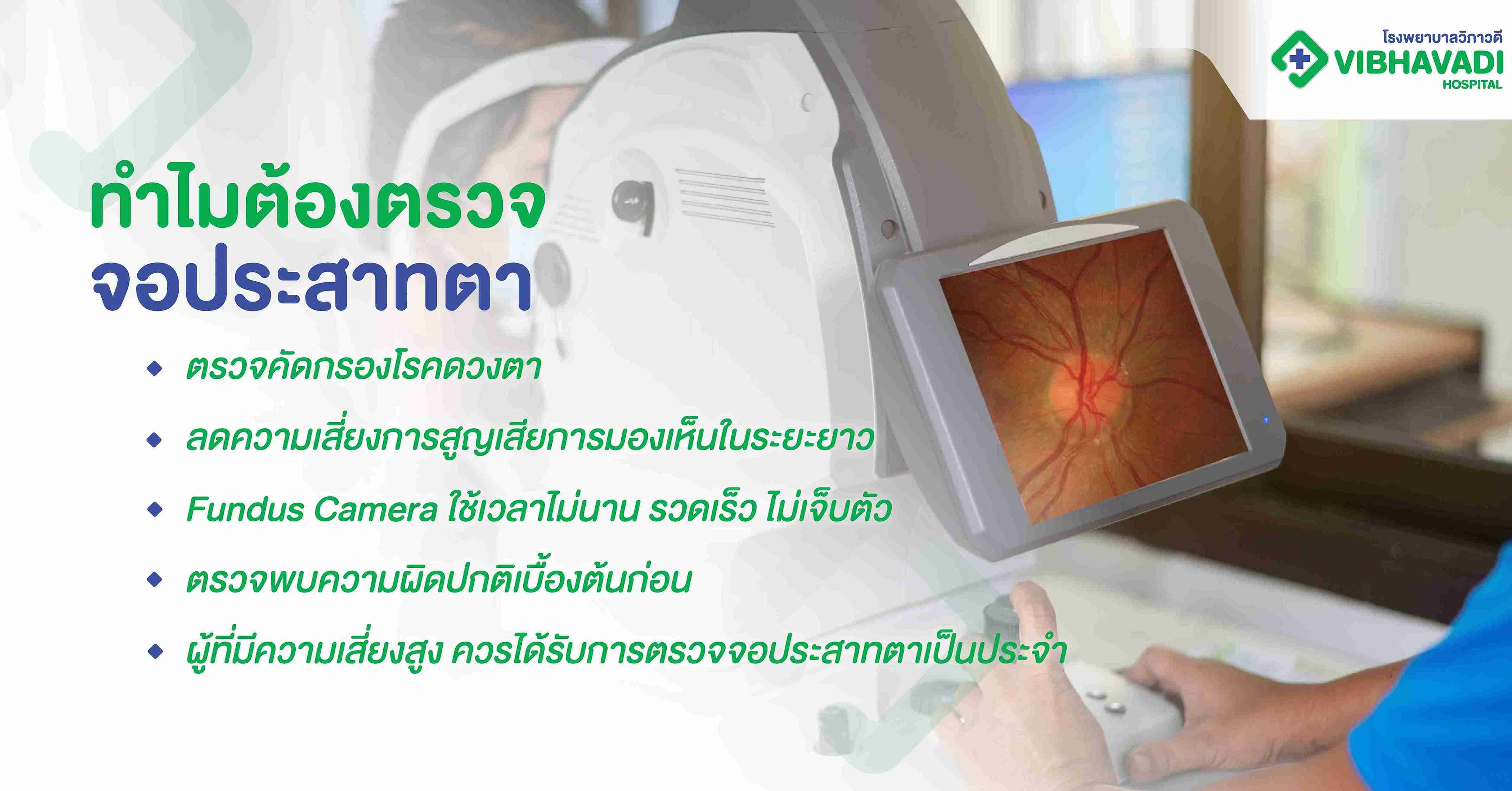
A fundus camera eye checkup is a non-invasive diagnostic procedure that captures high-resolution images of the interior surface of the eye, including the retina, optic disc, macula, and posterior pole. At Vibhavadi Hospital, we provide advanced retinal imaging services using state-of-the-art fundus camera technology. This allows for early detection of vision-threatening diseases such as diabetic retinopathy, glaucoma, macular degeneration, and hypertensive retinopathy.
What Is a Fundus Camera?
A fundus camera is a specialized low-power microscope with an attached camera designed to photograph the interior surface of the eye (the fundus). The images it captures are used by ophthalmologists to detect and monitor a wide range of eye conditions and systemic diseases that affect the retina and optic nerve.
Why Retinal Imaging Matters
The Importance of the Retina
The retina is a light-sensitive tissue lining the back of the eye. It plays a critical role in vision by converting light into neural signals sent to the brain. Retinal damage is often irreversible and can lead to partial or complete vision loss if not detected early.
Silent Progression of Eye Diseases
Many serious eye diseases develop without noticeable symptoms in the early stages. Regular retinal imaging allows for:
-
Early diagnosis
-
Timely intervention
-
Monitoring disease progression
-
Preventing vision loss
Who Should Get a Fundus Camera Eye Checkup?
High-Risk Groups
The following individuals are strongly recommended to undergo a fundus camera eye checkup:
-
Diabetics (Type 1 and Type 2)
-
Hypertensive patients
-
Individuals over 40 years old
-
Smokers and heavy alcohol consumers
-
People with a family history of glaucoma, AMD, or retinal disorders
-
Patients with frequent migraines or visual aura
-
Individuals with high cholesterol or cardiovascular risk
People With These Symptoms Should Also Consider Screening:
-
Blurred or distorted vision
-
Difficulty seeing at night
-
Flashes of light or floaters
-
Sudden vision loss
-
Eye pain or pressure
Common Conditions Detected by Fundus Photography
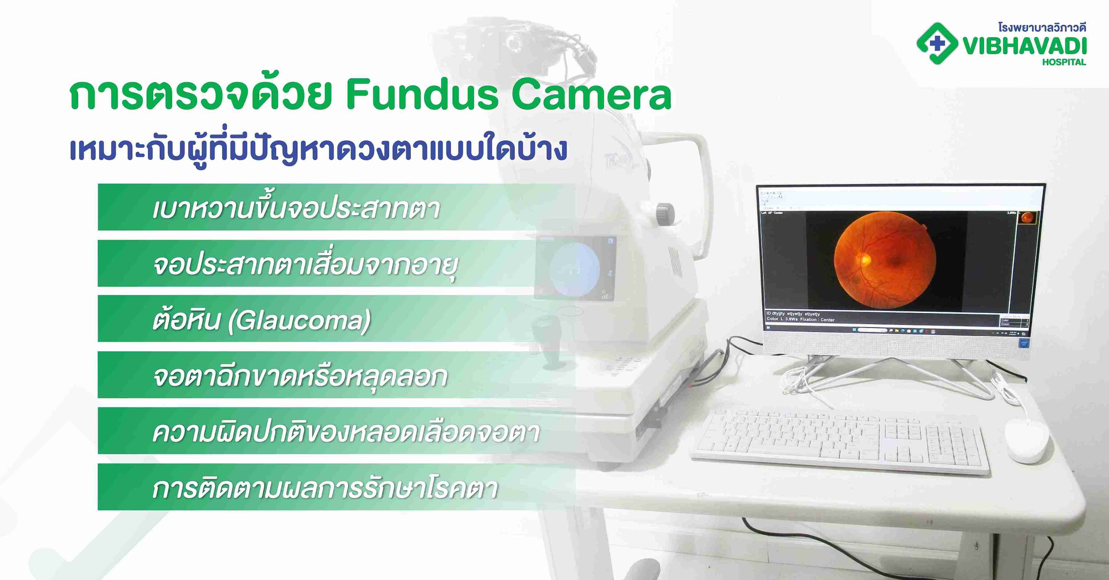
Diabetic Retinopathy
A common complication of diabetes, this condition damages the blood vessels in the retina and can lead to blindness if untreated.
Glaucoma
Increased intraocular pressure can damage the optic nerve, causing gradual vision loss.
Age-related Macular Degeneration (AMD)
This affects central vision, often in adults over 60, and is a leading cause of blindness.
Hypertensive Retinopathy
High blood pressure can cause structural damage to the retina, leading to blurred vision or even bleeding within the eye.
Retinal Detachment or Tears
These require urgent care and can be visually assessed with fundus imaging.
How the Fundus Camera Procedure Works
Step-by-Step Process
-
Pupil Dilation
You may be given eye drops to dilate your pupils for better visibility of the retina. -
Image Capture
You will rest your chin on a support while the camera takes images of the back of your eye. -
Non-Invasive and Quick
The procedure is painless, takes only 5–10 minutes, and requires no downtime.
No Preparation Required
There is no special preparation needed. However, if dilation drops are used, you should avoid driving for a few hours post-exam.
Benefits of Fundus Camera Eye Checkup
-
Non-invasive and painless
-
Quick and efficient
-
Highly detailed imaging for accurate diagnosis
-
Can be stored and used for future comparison
-
Supports early intervention in eye and systemic diseases
Vibhavadi Hospital's Retinal Imaging Services
Our Advanced Fundus Camera Technology
We use high-resolution digital fundus cameras capable of wide-angle and detailed imaging. Our equipment enables:
-
Fluorescein angiography (optional)
-
Optical coherence tomography (OCT) integration
-
Automated and manual capture modes
-
Secure image storage for tracking patient history
Professional Interpretation by Expert Ophthalmologists
Every scan is reviewed by our board-certified ophthalmologists, trained in both retinal diseases and systemic conditions visible through fundus imaging. Detailed reports and recommendations are provided promptly.
Seamless Referral for Further Care
If the scan reveals abnormalities, patients are referred to specialists such as:
-
Retina specialists
-
Glaucoma specialists
-
Internal medicine or endocrinology (for diabetes or hypertension)
Related Services at Vibhavadi Hospital
-
Comprehensive eye exams
-
Diabetic eye screening
-
Glaucoma screening and management
-
Macular degeneration treatment
-
OCT (Optical Coherence Tomography)
-
IOP (Intraocular Pressure) testing
-
LASIK and ICL surgery consultation
Booking a Fundus Camera Checkup
How to Make an Appointment
You can schedule your checkup via:
-
Website: https://www.vibhavadi.com/th/package/fundus-camera-eye-checkup
-
Call Center: Contact the Eye Center for available slots
-
Walk-in: Visit our Ophthalmology Department on-site
Clinic Hours
-
Monday–Friday: 8:00 AM – 5:00 PM
-
Saturday: 9:00 AM – 12:00 PM
-
Closed on Sundays and public holidays
Cost and Coverage
Checkup Package Price
The Fundus Camera Eye Checkup at Vibhavadi Hospital is currently offered at:
Only 600 THB (subject to hospital promotions)
This includes:
-
Fundus image capture
-
Expert interpretation
-
Preliminary consultation
Insurance and Health Benefits
-
Covered under some corporate health screening programs
-
Tax-deductible as health checkup expense in Thailand
-
May be claimable through private insurance (check with provider)
Meet Our Medical Team
Our Ophthalmology Team includes:
-
Retinal and macular disease specialists
-
Diabetic eye care experts
-
Glaucoma management professionals
-
Experienced technicians trained in advanced imaging systems
Each member is committed to high-standard, patient-centered care.
Frequently Asked Questions (FAQ)
Is the fundus camera procedure safe?
Yes. It is a non-invasive, completely safe procedure that uses light and digital imaging—not X-rays or radiation.
Will it hurt?
Not at all. The procedure is quick and painless, although dilation drops might cause temporary light sensitivity.
Do I need this checkup even if I have no vision problems?
Yes. Many retinal conditions, especially diabetic and hypertensive retinopathy, do not show symptoms until advanced stages.
How often should I get this checkup?
-
Diabetics: Annually or as advised
-
Individuals over 40: Every 1–2 years
-
Anyone with vision symptoms: Immediately
Can children have a fundus camera exam?
Yes, particularly if they have systemic diseases or symptoms. Pediatric retinal screening is available upon request.