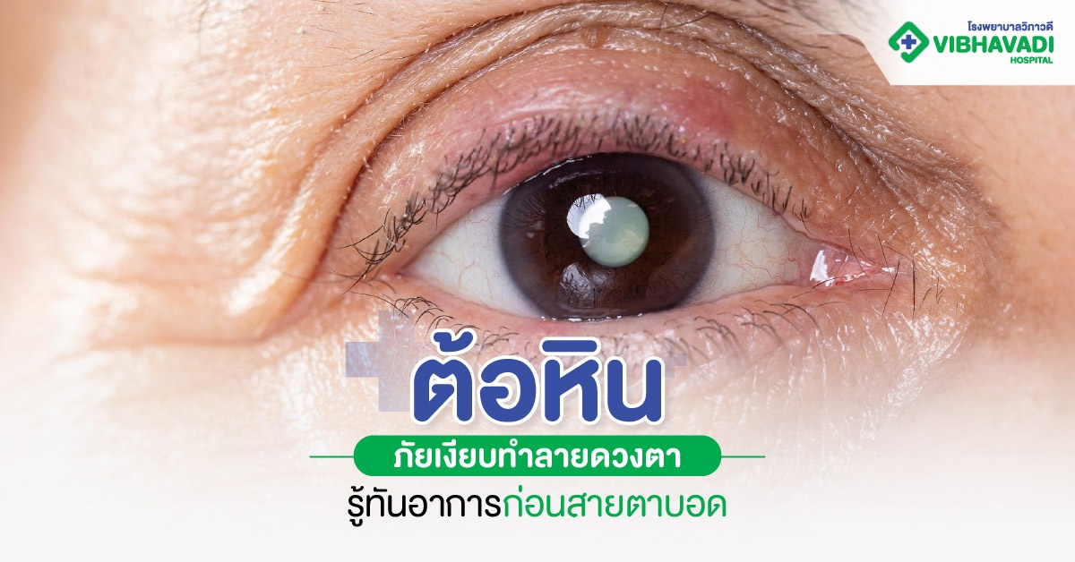
Glaucoma: Silent Eye Threat

Glaucoma: The Silent Threat That Destroys Vision – Recognize Symptoms Before Blindness Occurs
Key Takeaway
-
Glaucoma is a disease that damages the optic nerve due to high intraocular pressure (IOP). If left untreated, it can lead to permanent vision loss.
-
Early symptoms of glaucoma are often subtle, such as narrowing peripheral vision or blurriness. Some types may cause acute eye pain or seeing rainbow halos around lights.
-
Glaucoma treatment includes various methods like using medication to lower IOP, laser therapy, and surgery, depending on the type and severity of the disease.
"Glaucoma," often called the silent thief of sight, is an overlooked eye threat. It results from elevated pressure inside the eye, slowly damaging the optic nerve. In the early stages, it often shows no symptoms. If left undiagnosed and untreated, it can lead to blurred vision and eventually permanent vision loss. Regular eye exams and recognizing the warning signs are therefore the key to keeping your sight safe.
What is Glaucoma?
Glaucoma is caused by high intraocular pressure (IOP) or the optic nerve being overly sensitive to pressure, leading to optic nerve damage. This results in the visual field gradually narrowing. Without treatment, it can lead to permanent vision loss or blindness. The key characteristic is the slow, progressive degeneration of the optic nerve. Because it often presents with no obvious symptoms in the early stage, it’s nicknamed the "silent killer of sight."
Glaucoma is divided into several types, including:
-
Open-Angle Glaucoma
-
Angle-Closure Glaucoma
-
Congenital Glaucoma
-
Secondary Glaucoma
Each type has different severity and treatment guidelines, making regular eye check-ups essential to prevent long-term vision loss.
What Causes Glaucoma?
Glaucoma can stem from multiple causes and isn't solely due to high eye pressure. Other factors can accelerate optic nerve deterioration, increasing the risk of vision loss. Key factors include:
-
Excessively high intraocular pressure that presses on and damages the optic nerve's function.
-
Genetic factors: Having a family member with glaucoma increases your risk.
-
Age: It's more common in people over 40 as eye tissues naturally degenerate with age.
-
Underlying health conditions: Such as diabetes and high blood pressure, which affect blood flow to the optic nerve.
-
Use of certain medications: Especially long-term steroid use, which can increase eye pressure.
-
Eye injuries or other eye conditions: Such as trauma to the eye or other types of cataracts that affect fluid drainage within the eye.
Check Glaucoma Symptoms Before Vision Loss Occurs
Glaucoma is dangerous because it often shows no clear symptoms initially. Many confuse the mild blurred vision of early glaucoma with eye strain or general refractive errors. The difference is that normal blurry vision improves with rest or correct glasses, but if it's glaucoma, the symptoms will progressively worsen and may lead to permanent vision loss. Let's look at the symptoms for each type of glaucoma:
Open-Angle Glaucoma
This is the most common type, caused by the slow blockage of the eye's drainage system, leading to a gradual rise in IOP. It is often asymptomatic in the early stages. Vision may begin to narrow peripherally, eventually leaving only central vision, like looking through a tube (tunnel vision). It is common in older adults and those with a family history of glaucoma.
Angle-Closure Glaucoma
This type occurs when the eye's drainage angle is suddenly blocked, causing a rapid, acute rise in IOP. Symptoms often include sudden, severe eye pain, eye redness, seeing rainbow halos around lights, nausea, vomiting, and immediate blurred vision. It's typically found in people with farsightedness or deep-set eyes, especially middle-aged women and older.
Congenital Glaucoma
Congenital glaucoma is usually caused by abnormalities in the eye's drainage system present from birth. Symptoms include the child having an abnormally large eye size (Bupthalmos), excessive tearing, light sensitivity (photophobia), and a cloudy cornea. It is found in infants from birth or within the first years of life.
Secondary Glaucoma
Secondary glaucoma is caused by other factors affecting eye pressure, such as inflammation, injury, long-term steroid use, or certain eye diseases. Symptoms may include eye redness, pain, blurred vision, or partial vision loss. It's common in patients with chronic eye diseases, a history of eye injury, or those on certain medications long-term.
Which Glaucoma Symptoms Warrant a Doctor Visit?
Some glaucoma symptoms may seem like general vision issues, but leaving them untreated can cause permanent vision loss. You should see a doctor immediately if you experience the following:
-
Abnormal vision, such as seeing halos around lights (light scatter) or double vision.
-
Narrowing visual field, seeing less on the sides, as if looking through a tube.
-
Severe eye pain, especially combined with eye redness and sudden blurred vision.
-
Nausea and vomiting accompanying eye pain and blurred vision.
-
In small children, abnormally large eyes, excessive tearing, or a cloudy cornea should raise suspicion of congenital glaucoma.
If you have any abnormal eye symptoms, especially acute eye pain, sudden blurring of vision, or a noticeable change in your sight, you must see an ophthalmologist immediately to prevent irreversible damage to the optic nerve.
Methods for Diagnosing Glaucoma
Glaucoma diagnosis is not a simple vision test; it involves a comprehensive eye health check to look for abnormalities in intraocular pressure, the optic nerve, and the visual field. Early detection allows for disease control before permanent damage occurs. Diagnostic methods include:
Tonometry (Intraocular Pressure Test)
This is the basic method used to screen for glaucoma. The device measures the pressure inside the eyeball. The normal average value is between 10-21 mmHg. A higher value may indicate a risk of glaucoma, but not everyone with high IOP has glaucoma, so other tests are used in conjunction.
Visual Field Test
This test checks peripheral or side vision. The patient sits, focusing on a central point, and small lights appear at various spots. The patient presses a button when they see the light. This test checks for any loss in the visual field, a characteristic sign of glaucoma. In the early stages, patients may not realize their visual field has narrowed.
Gonioscopy (Test of the Angle Between Cornea and Iris)
The doctor places a small, mirrored lens on the eye (after applying anesthetic drops) to examine the angle between the cornea and the iris to see if it is open or blocked. This is crucial for distinguishing between types of glaucoma, such as Open-Angle Glaucoma (fluid drains slowly but the angle is open) and Angle-Closure Glaucoma (the angle is blocked, preventing drainage).
Additional Testing
In addition to the basic tests, the doctor may recommend supplementary tests for a more accurate diagnosis, such as:
-
OCT (Optical Coherence Tomography): Detailed imaging of the optic nerve to assess the degeneration of nerve fibers.
-
Pachymetry: Measurement of corneal thickness, as the thickness affects the measured IOP value.
-
Fundus Examination: Examination of the retina to check the shape of the optic nerve head for damage caused by glaucoma.
Glaucoma Treatment Guidelines
Glaucoma treatment varies depending on the type, severity, and the patient's overall eye health. The main goal is to lower intraocular pressure, prevent further optic nerve damage, and maintain stable vision.
Medication
Medication is the most common treatment, especially for those with mildly elevated IOP without severe damage. Eye drops must be used regularly as prescribed by the doctor to directly lower IOP. Medications either reduce the production of fluid in the eye or increase its outflow. Consistent use can slow down optic nerve damage, but side effects like dry eyes, redness, or irritation should be monitored. Some types may also affect heart or blood pressure, requiring consultation with a doctor before use.
Laser Therapy
Laser treatment is suitable for certain groups, such as Angle-Closure Glaucoma or some cases of Open-Angle Glaucoma. The doctor uses laser energy to create a new drainage path or improve fluid circulation in the eye, helping to lower eye pressure. This method is often a one-time procedure with noticeable results and is quick, but follow-up checks are necessary as the treatment effect may gradually decrease over time.
Surgery
Glaucoma surgery creates a new drainage path for the eye to permanently lower pressure. Methods include Trabeculectomy, which creates a temporary outflow channel beneath the eye's surface, or implanting a Glaucoma Drainage Device. Close post-operative eye care is necessary to prevent infection and complications. Strict adherence to the doctor's instructions is crucial for treatment success.
Post-Treatment Follow-up
Glaucoma treatment is not complete after medication, laser, or surgery, as the disease can recur. Regular follow-up checks allow the doctor to evaluate intraocular pressure, visual field, and the optic nerve to adjust the treatment plan accordingly and keep vision stable for longer, enabling the patient to live safely.
Guidelines for Glaucoma Care and Prevention
Glaucoma prevention starts with regular overall eye and health care. Since glaucoma often has no obvious symptoms in the early stages, the following care practices can help reduce the risk of various eye diseases:
-
Regularly visit an ophthalmologist for eye exams, especially if you are over 40 or have a family history of glaucoma.
-
Manage general health by controlling underlying conditions like diabetes, high blood pressure, and heart disease. Regular exercise also improves blood circulation to the eyes, reducing the risk of high IOP.
-
Avoid eye risk factors like unnecessary steroid use, avoiding trauma or impact to the eyes, and resting your eyes when performing visually demanding tasks to help reduce glaucoma risk.
-
Be aware of abnormal eye symptoms, such as blurred vision, eye pain, seeing rainbow halos, a narrowing visual field, or abnormally large eyes in children. If these symptoms occur, see an ophthalmologist immediately.
Glaucoma Treatment at Viphavadi Hospital
The Ophthalmology Center at Viphavadi Hospital offers comprehensive glaucoma screening and diagnosis, starting with intraocular pressure checks, visual field testing, and optic nerve examinations to diagnose the disease in its early stages.
For treatment, Viphavadi Hospital provides a full range of services, including eye drops to lower IOP, laser therapy to improve fluid drainage, and surgery for patients needing permanent IOP control. Furthermore, continuous post-treatment follow-up is provided to prevent vision loss and maintain stable eye health, ensuring your sight receives optimal care.
Summary
Glaucoma is a disease that damages the optic nerve due to high IOP. Without treatment, it can lead to permanent vision loss. Symptoms are often subtle early on, such as a narrowing visual field, or acute pain in some types. Glaucoma is classified as open-angle, angle-closure, congenital, and secondary. Diagnosis involves testing intraocular pressure, the visual field, the corneal angle, and further tests like OCT. Treatment covers medication, laser, and surgery, along with regular follow-up, to reduce the risk of vision loss. Prevention is achieved through regular eye exams, health management, avoiding risk factors, and monitoring for abnormal symptoms.
Get Glaucoma Tested and Treated—a disease that destroys the optic nerve and can cause permanent vision loss if untreated. The Ophthalmology Center at Viphavadi Hospital offers comprehensive screening and treatment for glaucoma, including medication, laser, and surgery, with experienced ophthalmologists and modern technology. We provide professional care for all eye problems. Interested in testing or treatment? Find more information at Viphavadi Hospital or call 02-561-1111.
Frequently Asked Questions (FAQ)
Understanding glaucoma helps in managing the disease correctly and reducing the risk of vision loss.
Can Glaucoma be Cured?
Glaucoma cannot be completely cured, but intraocular pressure can be controlled, and optic nerve damage can be slowed down with appropriate treatment, such as eye drops, laser, or surgery. Regular follow-up helps prevent permanent vision loss.
What Symptoms Require Glaucoma Surgery?
Patients may need surgery if medication and laser treatments fail to control the intraocular pressure or if there is clear damage to the optic nerve. Surgery helps create a new drainage path, lowering IOP and preventing permanent vision loss.
Is Glaucoma Laser Treatment Painful?
Glaucoma laser treatment is generally not severely painful. The doctor will apply anesthetic drops to reduce sensation. The patient may feel only mild warmth or pressure during the procedure, which is completed quickly, allowing the patient to go home afterward.
How Long is the Recovery Time for Glaucoma Surgery?
After surgery, most patients can return to their normal daily life within 1-2 weeks, depending on the type of surgery and individual recovery. The doctor will schedule close follow-up appointments to monitor intraocular pressure and vision.
Testimonials
Proud to take care of you


